Posterolateral Lumbar Fusion
Stabilizing selected lumbar spinal segments by encouraging new bone to grow between adjacent vertebrae outside the disc space.
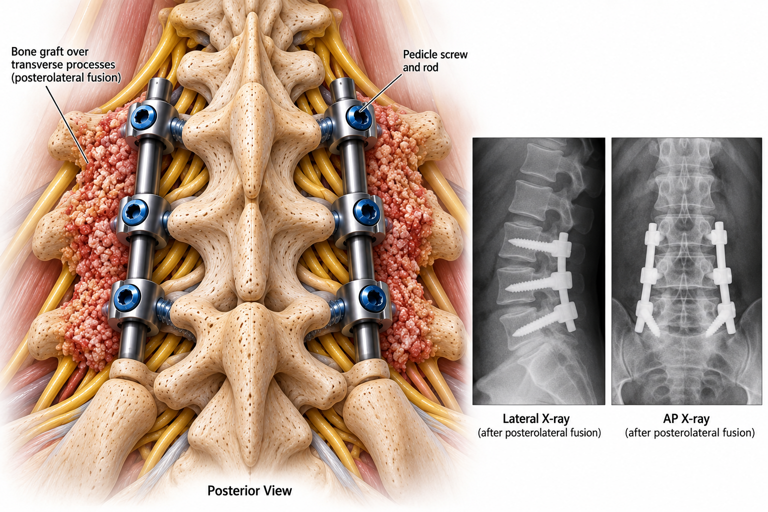
What is posterolateral lumbar fusion?
Posterolateral lumbar fusion is a surgical technique intended to join two or more vertebrae into one stable segment. Rather than placing a spacer into the disc space, the surgeon places bone graft along the back and sides of the spine, typically over the transverse processes and adjacent posterolateral bony surfaces. Over time, the graft may incorporate and form a continuous bridge of bone between the vertebrae.
How is the fusion performed?
After exposing the appropriate lumbar levels, the surgeon identifies the transverse processes and other posterolateral bony surfaces. These surfaces are prepared, or decorticated, to expose bone that can support healing. Bone graft is then placed across the prepared region to encourage a solid fusion mass. Depending on the condition, the graft may include the patient's own bone, donor bone, or other bone-graft materials.
Pedicle screws are often placed through the pedicles into the vertebral bodies and connected with rods. This instrumentation provides immediate mechanical stability while the graft heals; the screws and rods do not themselves constitute a completed bony fusion. If nerves are compressed, decompression may be performed during the same operation when indicated.
How is this different from TLIF?
In a transforaminal lumbar interbody fusion (TLIF), a spacer and bone graft are placed into the disc space between vertebral bodies. In a posterolateral fusion, graft is placed outside the disc space along the transverse processes. In some operations, surgeons combine an interbody fusion with posterolateral bone grafting. The choice depends on spinal alignment, instability, nerve compression, prior surgery, and other individual factors.
When might it be considered?
Lumbar fusion may be considered for selected patients with spinal instability, certain forms of spondylolisthesis, deformity, or other conditions when stabilization is needed. Surgery is not automatically indicated for degenerative changes or back pain alone; imaging, symptoms, examination findings, and response to nonoperative treatment help guide the decision.
Recovery and potential risks
Fusion is a biological healing process that usually takes months. Follow-up visits and imaging may be used to assess alignment, hardware, and progression of healing. Risks can include infection, bleeding, dural tear, nerve injury, blood clots, failure of the bones to fuse (nonunion), hardware complications, and degeneration at adjacent levels. Individual recovery recommendations depend on the procedure and the patient's health.